An ultrasonic scan creates a picture of a person’s inner physiological systems by using high-frequency sound waves to travel through the body. In humans, it is commonly used to evaluate the developing foetus, the abdominal and pelvic organs, the muscles, and tendons, as well as the heart and blood vessels of the individual. An ultrasound scan is sometimes referred to as a sonogram or an echocardiogram in some circles (when imaging the heart).
Transmitted high-frequency sound waves are directed at the inside body structures that are being probed by the ultrasound equipment, which is a type of medical imaging technology. An image is created by recording the echoes or reflected noises, which is then shown on a computer monitor or television. To transmit and receive sound waves, a small, hand-held probe is employed to do so. Because of the high frequency of the sound, the human ear is unable to detect it, which is why it is referred to as ultrasonic.
Most of the time, a portable ultrasound machine scan is non-invasive (done from outside the body). For some scans, a particular probe must be inserted into the patient’s vagina for some obstetric or pelvic examinations, into the rectum for some prostate examinations, or into the oesophagus for some cardiac examinations. The same is true for dogs who have veterinary ultrasonography examinations. Ultrasound scanning may be used by doctors to monitor and guide invasive procedures such as a biopsy of a person’s breast or thyroid gland, among other things.
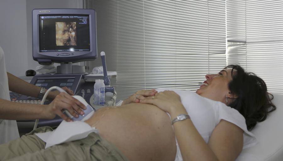
Foetal anomalies, such as spina bifida, are detected by pregnancy and pelvic scans, which are also used to assess the age and location of a foetus, as well as track the development of the foetus during pregnancy.
Additional uses include musculoskeletal scans of the shoulder, hip, and elbow, as well as breast scans, among others. Consider a scan of a person’s eye to analyse its inner structures and to further investigate an aberration discovered through physical examination or mammography, for example.
Known as Doppler ultrasonography, this type of ultrasound measures the speed and direction of blood flow in specific parts of the body, such as the neck arteries and leg veins, using high-frequency sound waves.

What is the procedure for performing an ultrasound technique?
When you have an upper abdomen ultrasound, you will need to lie down on an examining table or in a bed. A gel will be applied to your skin by the ultrasound technician, who is also known as a sonographer, to aid the ultrasonic probe in making better contact with your body.
On a computer monitor, real-time representations of the two-dimensional (and occasionally three-dimensional) images are displayed. Other ultrasounds may necessitate the use of a slightly different method. A transvaginal scan may be performed on a woman who is having her pelvic evaluated. This procedure involves inserting a specific ultrasonic probe into the vagina rather than, or in addition to, scanning the pelvis through the front of the pelvis. According to the information provided, the necessity for ultrasound is extremely significant, particularly in the case of women.